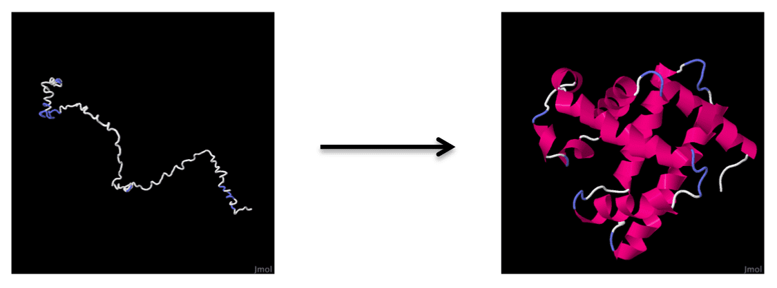
Helix Formation – Myoglobin
Click the structures and reaction black arrows in sequence to view the 3D models and animations respectively

The animation is simply based on the results of a series of calculations for rational intermediate frames using the Yale Morph Server and Spartan08 Fixer to produce viable pdb files. It is a rough demonstration of protein folding.
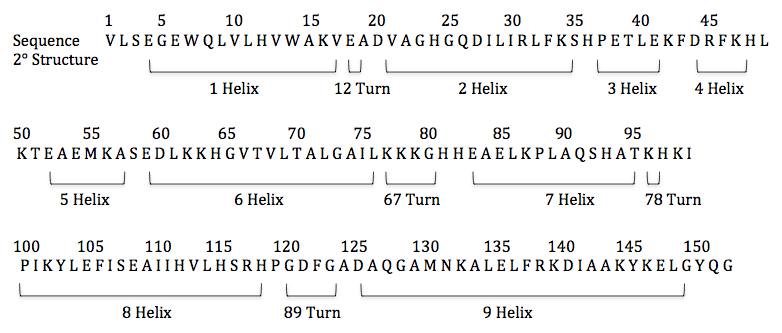
Experimentally, the first step of folding myoglobin is found to be the formation of secondary structures of 1, 7 and 8 helices (shown below), followed by 2 helix and then the rest of the protein. Native secondary structures are expected to form in their original regions and assemble into final structure by collision and diffusion.

The sequence of Myoglobin:

Framework folding | Hairpin folding | Myoglobin helix formation


