Please note, due to the complexity of the structure this page may take longer to load
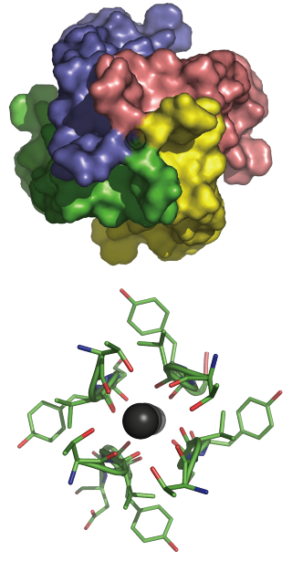
Top: View of the enzyme from inside the cell showing the entrance pore that admits hydrated ions.
Bottom: View looking up the selectivity filter showing how mobile dehydrated K+ ions are coordinated by peptide carbonyl O atoms provided by each of the four subunits. Note the almost four-fold symmetry axis.